

Stomach fluke (paramphistomes) in ruminants
By Dr Joan Lloyd, Dr Joe Boray, and Stephen Love Veterinarian/State Worm Control Coordinator, Extensive Industries Development, Armidale and published by NSW Department of Primary Industries - This Primefact looks at stomach fluke (paramphistomes) parasites of ruminants which particularly affect cattle and sheep.The flukes are often found in small numbers but only seriously affect livestock under certain conditions, in heavy infections, and in certain growth stages.
Adult flukes in the rumen (first stomach or paunch) or reticulum (second stomach or honeycomb), for instance, are not known to cause clinical disease in Australia. However, damage to the rumen due to heavy infections has been recorded.
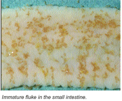
Heavy infections with immature flukes in the upper small intestine can cause serious ill-health and death.
Life cycle
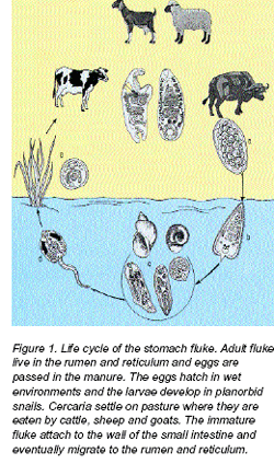
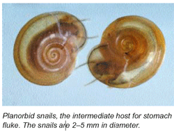
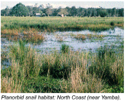
The stomach fluke has a complex life cycle which requires an intermediate host for completion. The intermediate hosts in Australia are planorbid snails. These are small, flat snails from 2–5 mm in diameter.The two species of stomach fluke which infect ruminants in Australia, Calicophoron calicophorum and Paramphistomum ichikawai, are transmitted by the snails Gyraulus scottianus and Helicorbis australiensis, respectively.
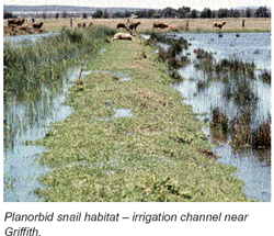
These snails are found in permanent and temporary watercourses, irrigation channels, swamps, dam edges and depressions. They are normally found attached to vegetation in these habitats.
Adult stomach flukes live in the rumen and reticulum of cattle, sheep and goats. The eggs are passed in manure. Larvae then hatch in a wet environment and infect the intermediate host – planorbid snails.
Larval development is completed in the snail, and the next stage, the cercariae, leave the snail and attaches to vegetation where it encysts (metacercariae).
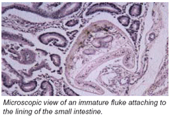
When the vegetation is grazed by ruminants, the immature fluke ex-cysts and attaches itself to the walls of the small intestine. It later migrates to the rumen and reticulum to become an egg-producing adult. In light infections, young flukes migrate to the rumen within 4–6 weeks and normally no clinical symptoms occur. Egg production begins soon after the fluke enter the rumen.
In heavy infections, development of the young flukes is retarded and they can stay in the small intestine for more than four months and cause severe disease associated with destruction of the lining of the small intestine. In a severe outbreak, up to 72,000 immature flukes may be present in the small intestine.
Distribution
Planorbid snails must be present in the local environment to complete the stomach fluke life cycle. This limits outbreaks of infection to areas where the snail is found. Light infections, due to adult and immature flukes, occur on the Southern and Central Tablelands, the Slopes, and in coastal areas. Serious outbreaks from heavy infections with immature flukes may occur on the New England Tablelands, the North and South Coast, and in irrigation areas.Clinical signs
Most livestock have only light stomach fluke infections. They show no signs of disease due to either adult fluke or small numbers of immature fluke. Heavy infections with the immature fluke may cause decreased appetite, listlessness and weight loss. Fluid, foul-smelling diarrhoea, dehydration and death may result from these infections. Moderate infections with the immature fluke may cause reduced weight gains or milk production, or ill-thrift.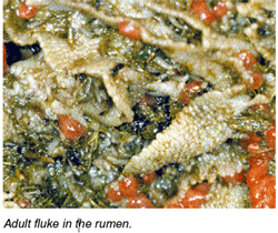
Immunity
Cattle, sheep and goats develop resistance after exposure to the parasite. This immunity protects the animal against the massive infections of immature fluke that cause the most problems.Weaner cattle and lambs appear to be the most susceptible.
Adult animals can still be affected, particularly if they have had no prior exposure to the parasite or are debilitated by other problems, such as unbalanced nutrition.
Diagnosis
Diagnosis relies on a combination of post mortem findings, history of the outbreak, clinical signs observed in the animal, and response to drenching. Other conditions can also be confused with stomach fluke disease. The most commonly mistaken conditions are roundworm and liver fluke infections in cattle and sheep, and copper deficiency in cattle. Various bacterial, viral and protozoal infections and poisoning by plants and chemicals can also cause similar signs. In view of the number of diagnostic possibilities, the assistance of a veterinarian should be sought in making a diagnosis.A careful examination of the small intestine with a magnifying glass or special microscope is required to confirm a diagnosis. The presence of eggs in manure only indicates the presence of adult fluke and has no diagnostic value for an outbreak of the acute disease.
Treatment
Adult stomach flukeAlthough treatment for adult fluke has no direct benefit to the animal, it may reduce the source of infection for the snail intermediate host. This then reduces the size of the next generation of infective fluke larvae on pasture. Treatment with an appropriate drench should be timed for autumn and spring.
Effective treatment of immature stomach fluke infection requires removal of stock from the source of infection, usually swampy land, as well as treatment with a drench which is effective against immature fluke. Supportive therapy to treat dehydration and any secondary infection may be needed. Seek veterinary advice before starting treatment.
Anthelmintic treatment There are currently no drenches registered in NSW for use against paramphistomes. The only way to legally undertake such treatment is by way of ‘off-label’ use under veterinary direction.
Veterinarians are referred to papers published by Rolfe and Boray (see ‘Further Reading’). Prescribing veterinarians will need to consider the residue implications of Rolfe and Boray’s recommended treatment regimen.
Control
Although there are practical limitations, the following measures provide some control of stomach fluke.- Drain affected areas.
- Fence off affected areas.
- Provide alternative water sources.
- Treat stock with drenches effective against stomach flukes.
Serious outbreaks may be prevented if pastures with a natural water supply are grazed only in spring and early summer. Late summer, autumn and early winter grazing should be avoided. If wet pastures are grazed continuously, young weaner calves and lambs should not be grazed together with adults.
The control that can be achieved by these methods will depend on the circumstances on an individual property. Contact your private veterinarian, district veterinarian or veterinary officer to formulate specific control strategies.
Further information
For further information, contact your veterinary practitioner, district veterinarian, or local NSW Department of Primary Industries office.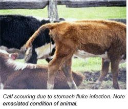
Further reading
Rolfe, PF and Boray, JC (1987). Chemotherapy of paramphistomosis in cattle. Australian Veterinary Journal 64:328-332.Rolfe PF and Boray JC (1988). Chemotherapy of paramphistomosis in sheep. Australian Veterinary Journal 65:148-150.
Rolfe PF, Boray JC, Nichols P and Collins GH (1991). Epidemiology of paramphistomosis in cattle. International Journal for Parasitology 21:813-819.
Rolfe PF, Boray JC and Collins GH (1994). Pathology of infection with Paramphistomum ichikawai in sheep. International Journal for Parasitology 24:995-1004.
Acknowledgement
This Primefact (a further revision by S. Love of Lloyd and Boray’s Agfact) is largely based on the second edition (1985) Agfact written by Dr Peter Rolfe.© State of New South Wales, Australia.

